Knee Arthroscopy

knee arthroscopy adelaide

Knee arthroscopy surgery to investigate and treat joint damage

Knee arthroscopy is commonly performed to diagnose and treat a wide range of knee problems, including cartilage damage, meniscal tears, loose bodies, inflammation, or early degenerative changes.


Through small keyhole incisions, specialised instruments can be used to perform precise repairs or clean-up of damaged tissue, often avoiding the need for open surgery. Recovery is typically faster and less painful than traditional approaches, although rehabilitation and activity modification are still important.

Knee arthroscopy may be recommended if non-surgical treatments such as physiotherapy, medication, or injections have not improved your symptoms. Dr George Awwad will help determine whether arthroscopy is suitable for your condition based on your symptoms, imaging results, and activity goals.

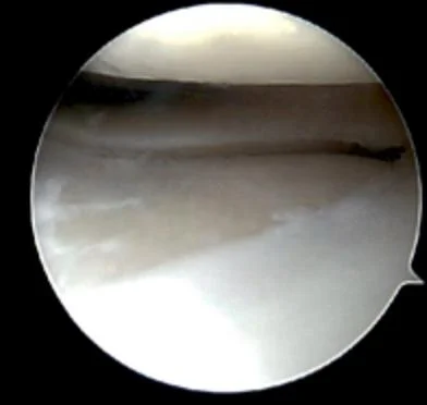
What is knee arthroscopy?

Knee arthroscopy is a minimally invasive surgical procedure used to diagnose and treat problems within the knee joint. It is commonly performed when imaging tests (such as X-rays or MRI) do not fully explain knee pain or when non-surgical treatments have not improved your symptoms.


During the procedure, Dr Awwad makes a few small incisions around the knee and inserts a tiny camera (arthroscope) to view the inside of the joint on a monitor. Specialised surgical instruments are then used to repair or remove damaged tissue as needed.

Knee arthroscopy can be used to treat a wide range of knee conditions, such as torn cartilage (meniscus), damaged ligaments, loose bone or cartilage fragments, inflamed joint linings, and patella (kneecap) tracking problems. Because the procedure is performed through small incisions, it generally involves less pain and a quicker recovery than traditional open surgery.


Dr Awwad will discuss whether knee arthroscopy is appropriate in your situation, based on your symptoms, imaging results, and overall knee health.

Conditions that may be treated with knee arthroscopy

Knee arthroscopy may be used to diagnose and treat a range of conditions that affect the structures within the joint. Some of the more common conditions treated using this minimally invasive procedure include:

1. Meniscal Tears

The menisci are two C-shaped pieces of cartilage that act as shock absorbers between the femur and tibia. Arthroscopy can be used to repair or remove torn meniscal tissue, depending on the type, location, and severity of the tear.

2. Loose Bodies

Fragments of bone or cartilage may break off and float within the joint, causing pain, swelling, or locking. These loose bodies can be located and removed arthroscopically.

3. Cartilage Damage

Articular cartilage injuries or defects can be smoothed, debrided, or treated with techniques such as microfracture to stimulate repair. This may help reduce joint pain and delay further degeneration.

4. ACL Injuries (Partial or Full Tears)

While full ACL reconstruction is typically performed through open or mini-open techniques, arthroscopy plays an important role in diagnosis and visual assessment of ACL injuries. It is also commonly used during ACL reconstruction to visualise the joint and guide graft placement.

5. Synovitis

Inflammation of the joint lining (synovium) may be addressed with arthroscopic synovectomy, particularly in cases of rheumatoid arthritis or other inflammatory joint disorders.

6. Patella Tracking Disorders

In cases of recurrent dislocations or maltracking of the kneecap, arthroscopy can be used to assess the patellofemoral joint and sometimes perform procedures to correct soft tissue imbalance.

7. Plica Syndrome

A thickened fold of the joint lining (known as a plica) can become inflamed and cause discomfort. Arthroscopic removal of the plica may relieve symptoms.

8. Early Arthritis and Joint Debridement

In patients with early-stage osteoarthritis, arthroscopy may be used to smooth damaged cartilage, flush the joint, or remove inflamed tissue. While not a cure, this may reduce pain in selected cases.

9. Baker’s Cyst (Secondary Treatment)

While not typically treated directly via arthroscopy, associated intra-articular pathology contributing to the formation of a Baker’s cyst (such as meniscal tears or arthritis) may be addressed.

When knee arthroscopy might be recommended

Knee arthroscopy may be recommended when non-surgical treatments such as physiotherapy, rest, medications, or injections have not resolved persistent knee symptoms. It is a minimally invasive option that allows your orthopaedic surgeon to both diagnose and treat issues inside the joint with reduced recovery time compared to open surgery.
Common reasons why knee arthroscopy may be recommended include:


Ongoing Knee Pain or Swelling

If you experience chronic knee pain or swelling that does not improve with conservative management, arthroscopy may help identify and treat the underlying issue.

Mechanical Symptoms (Locking, Catching, or Giving Way)

Symptoms such as the knee locking in place, catching during movement, or giving way unexpectedly may indicate internal joint problems such as loose bodies or meniscal tears that can be treated arthroscopically.

Suspected Cartilage or Meniscal Injury

If imaging (e.g. MRI or X-ray) reveals damage to the meniscus or articular cartilage, arthroscopy may be used to remove, repair, or smooth the affected tissue.

Limited Range of Motion

A reduced ability to fully bend or straighten the knee may result from internal joint damage, adhesions, or inflammation, which may be addressed through arthroscopy.

Unclear Diagnosis

In cases where imaging is inconclusive, but symptoms persist, diagnostic arthroscopy may be used to directly visualise the inside of the joint and guide further treatment.

Preparation for Further Surgery

Arthroscopy may be used as part of the planning process for more complex knee procedures, such as ligament reconstructions or osteotomies, especially where precise evaluation of the joint is needed. Dr Awwad will consider a range of factors including your symptoms, imaging results, overall health, and activity level—before recommending knee arthroscopy as a suitable treatment option.

Common arthroscopic knee procedures

Arthroscopic surgery is a versatile technique used to diagnose and treat a wide range of knee conditions. Below are some of the most common procedures performed arthroscopically:

Meniscus repair


Torn meniscus cartilage may be sutured or anchored back into place, particularly in younger patients or when the tear is located in a well-vascularised area of the meniscus.

In cases where the meniscus is not repairable, the damaged portion may be trimmed or removed to reduce pain and mechanical symptoms such as locking or catching.

A torn anterior cruciate ligament (ACL) is often reconstructed using a graft (typically from the hamstring or patellar tendon), restoring stability to the knee. Arthroscopy allows precise graft placement and minimal disruption to surrounding tissues.

Posterior cruciate ligament (PCL) injuries can also be managed arthroscopically using graft reconstruction techniques, often in conjunction with treatment for other ligament or cartilage injuries.

Loose fragments of bone, cartilage, or scar tissue may float within the knee joint and cause pain, swelling, or locking. Arthroscopy enables their safe and effective removal.

Areas of damaged cartilage may be smoothed (debridement), or, where appropriate, repaired or stimulated to promote healing, helping preserve the joint surface and reduce pain.

Selected intra-articular fractures of the knee may be assessed and in some cases treated arthroscopically, particularly if they involve the joint surface or require precise alignment.

For patients with early signs of osteoarthritis, arthroscopy may help relieve symptoms by removing inflamed tissue, smoothing cartilage, or addressing underlying causes such as a torn meniscus or impinging bone spurs.

Torn meniscus cartilage may be sutured or anchored back into place, particularly in younger patients or when the tear is located in a well-vascularised area of the meniscus.In cases where the meniscus is not repairable, the damaged portion may be trimmed or removed to reduce pain and mechanical symptoms such as locking or catching.A torn anterior cruciate ligament (ACL) is often reconstructed using a graft (typically from the hamstring or patellar tendon), restoring stability to the knee. Arthroscopy allows precise graft placement and minimal disruption to surrounding tissues.
Posterior cruciate ligament (PCL) injuries can also be managed arthroscopically using graft reconstruction techniques, often in conjunction with treatment for other ligament or cartilage injuries.Loose fragments of bone, cartilage, or scar tissue may float within the knee joint and cause pain, swelling, or locking. Arthroscopy enables their safe and effective removal.Areas of damaged cartilage may be smoothed (debridement), or, where appropriate, repaired or stimulated to promote healing, helping preserve the joint surface and reduce pain.Selected intra-articular fractures of the knee may be assessed and in some cases treated arthroscopically, particularly if they involve the joint surface or require precise alignment.For patients with early signs of osteoarthritis, arthroscopy may help relieve symptoms by removing inflamed tissue, smoothing cartilage, or addressing underlying causes such as a torn meniscus or impinging bone spurs.

The knee arthroscopy procedure: step-by-step

Knee arthroscopy is typically performed as a day procedure under general or regional anaesthesia. The following steps outline how the surgery usually unfolds:


1. Pre-operative preparation

Before the procedure begins, your knee will be cleaned with antiseptic solution and sterile drapes will be applied. You’ll be positioned on the operating table in a way that allows full access to your knee joint. A tourniquet may be placed on your thigh to control bleeding during the procedure.

2. Small incisions are made

Two or more small (approximately 1cm) incisions are made at the front or side of the knee. One is used to insert the arthroscope (a small camera), and the others allow insertion of specialised instruments.

3. Insertion of the arthroscope

A small camera connected to a light source and video monitor is inserted through one of the incisions. This allows the surgeon to inspect the inside of the knee joint in detail.

4. Joint inspection and diagnosis

The surgeon will systematically examine the cartilage, ligaments (such as the ACL and PCL), menisci, and the lining of the joint. Any abnormalities such as tears, loose bodies, inflammation, or degeneration are identified.

5. Treatment of identified issues

Once a diagnosis is confirmed, the surgeon may proceed to treat the condition during the same procedure. Common treatments include:


  • Trimming or repairing a torn meniscus

  • Removing loose fragments of bone or cartilage

  • Smoothing damaged cartilage (chondroplasty or debridement)

  • Reconstructing torn ligaments (e.g. ACL or PCL)

  • Synovectomy (removal of inflamed joint lining)

Specialised instruments such as shavers, graspers, or radiofrequency probes are used to carry out these treatments.

6. Flushing and closure

At the end of the procedure, the knee joint is flushed with sterile fluid to remove debris or blood. The instruments and arthroscope are removed, and the incisions are closed with sutures or adhesive strips.

7. Dressing and recovery

A sterile dressing is applied, and a compression bandage may be used to help minimise swelling. Patients are taken to recovery and typically discharged the same day with instructions for pain management, wound care, and follow-up.

Recovery and rehabilitation after knee arthroscopy

Recovery following knee arthroscopy is generally quicker than open knee surgery, but it still requires careful rehabilitation to ensure optimal healing and function. Dr Awwad’s post-operative protocol provides structured guidance for managing the recovery process in stages.


Immediately After Surgery


  • Hospital Stay: Most patients can go home on the same day of surgery.

  • Crutches: You may need crutches for support during the initial 24–72 hours, depending on your procedure and comfort.

  • Pain Management: Ice packs, elevation, and prescribed medications may help reduce swelling and manage discomfort.

  • Wound Care: Waterproof dressings allow showering but, these should remain clean and intact for the first 2 weeks.

Weeks 1–2: Early Mobility and Pain Control


  • Weight-Bearing: You will usually be allowed to bear weight as tolerated unless instructed otherwise for more complex procedures like meniscal repair.

  • Exercise Goals:

  • Regain full knee extension (straightening).

  • Initiate gentle range-of-motion (ROM) exercises and quadriceps activation (e.g. quadriceps sets and straight leg raises).

  • Precautions:

  • Avoid prolonged standing or walking.

  • Limit stairs and deep squats during this period. Avoid deep squats and uneven ground.

Weeks 3–6: Regaining Range and Strength


  • Activities:

  • Begin stationary bike (with no resistance) and pool walking (once wounds are fully healed).

  • Increase ROM exercises to aim for 0–120 degrees of flexion.

  • Add leg presses, mini squats, and hamstring curls as tolerated.

  • Physiotherapy: Regular sessions to guide progression, monitor swelling, and refine gait.

Weeks 6–12: Progressive Strengthening


  • Goals:

  • Achieve full range of motion.

  • Improve muscular control and balance.

  • Return to low-impact activities (e.g. elliptical trainer, gentle cycling).

  • Exercise Progression:

  • Introduce resistance training, proprioception, and closed-chain strengthening exercises.

  • Avoid pivoting, twisting, or impact activities unless cleared by your physiotherapist or surgeon.

Beyond 3 Months: Return to Sport or Work


  • Return to Sport: Gradual return to higher-level or sport-specific training, usually after 3 months depending on the extent of the procedure (e.g. meniscus repair vs. simple debridement).

  • Work: Most desk-based workers return within 1–2 weeks. Manual labourers may require 6–12 weeks or longer.

  • Ongoing Rehab: Home-based and/or gym exercises may be continued with physiotherapist oversight.

Follow-Up Appointments


  • You will typically see Dr Awwad for review within 1–2 weeks post-surgery to assess wound healing and range of motion.

  • Further follow-ups will monitor your functional progress and discuss return to activity timelines.

If you have any concerns during your recovery, including increased pain, redness, swelling, or signs of infection, please contact Dr Awwad’s rooms for prompt review.

Benefits of knee arthroscopy

Knee arthroscopy offers several important advantages over traditional open surgery, particularly for patients with ligament, cartilage, or meniscal injuries that require diagnosis or treatment. Some of the key benefits include:

Minimally invasive technique

Arthroscopy is performed through small incisions using a specialised camera and instruments, resulting in significantly less disruption to surrounding tissue compared to open surgery.

Reduced pain and swelling

Because the procedure is less invasive, patients typically experience less post-operative pain and swelling, allowing for a more comfortable recovery process.

Faster recovery and return to activity

Many patients can return to light activities within a few days and progress to more active rehabilitation sooner than with open procedures. Recovery timelines vary depending on the condition treated and the complexity of the procedure.

Clear visualisation of the joint

Arthroscopy provides the surgeon with a magnified, high-definition view inside the knee joint, allowing for accurate diagnosis and precise treatment of multiple conditions during the same procedure.

Lower risk of infection

The smaller incisions used in arthroscopic surgery are associated with a lower risk of wound infection compared to larger open incisions.

Outpatient or short hospital stay

Many arthroscopic procedures are performed as day surgery, meaning patients can go home on the same day, reducing the cost and inconvenience of a hospital stay.

Versatility in treatment

Knee arthroscopy can be used for a wide range of procedures, including:


  • Meniscal repair or removal

  • Ligament reconstruction (e.g. ACL or PCL)

  • Cartilage repair or debridement

  • Removal of loose bodies

  • Synovial tissue removal

  • Treatment of mild to moderate arthritis (joint preservation)

As with any procedure, outcomes can vary depending on individual factors. During your consultation, Dr Awwad will discuss the potential benefits and limitations of arthroscopy in your specific situation.

Risks and potential complications of knee arthroscopy

Knee arthroscopy is considered a low-risk procedure, particularly when compared to open surgery. Like any surgical intervention however, it carries potential risks and complications.

Possible risks include:

Infection


Although rare, there is a small risk of developing an infection at the incision sites or within the joint.

Mild bleeding around the surgical sites or bruising can occasionally occur.

Some patients may experience stiffness or reduced range of motion following the procedure. This is usually temporary and improves with physiotherapy.

Post-operative swelling and discomfort are common and typically settle within a few days to weeks.

While uncommon, damage to nearby nerves or blood vessels may occur.

There is a very small risk of developing blood clots in the leg after surgery.

In some cases, symptoms may persist or recur if the underlying condition does not respond fully to arthroscopic treatment.

As with any procedure involving anaesthesia, there is a small risk of adverse reactions, which will be reviewed during your pre-operative assessment.

Although rare, there is a small risk of developing an infection at the incision sites or within the joint.
Mild bleeding around the surgical sites or bruising can occasionally occur.
Some patients may experience stiffness or reduced range of motion following the procedure. This is usually temporary and improves with physiotherapy.Post-operative swelling and discomfort are common and typically settle within a few days to weeks.While uncommon, damage to nearby nerves or blood vessels may occur.There is a very small risk of developing blood clots in the leg after surgery.In some cases, symptoms may persist or recur if the underlying condition does not respond fully to arthroscopic treatment.As with any procedure involving anaesthesia, there is a small risk of adverse reactions, which will be reviewed during your pre-operative assessment.

It’s important to note that the likelihood of serious complications is low, and many patients recover smoothly with appropriate post-operative care. Your individual risks will be discussed in more detail during your consultation with Dr Awwad, including how to prepare for surgery and minimise these risks.

FAQs about knee arthroscopy

What is knee arthroscopy used for?


Knee arthroscopy is a minimally invasive procedure used to diagnose and treat a range of knee problems. It may be used to assess joint damage, repair torn cartilage or ligaments, remove loose fragments, or address joint inflammation.

Most patients can walk with support within a day or two. Return to work and everyday activities may take 1–3 weeks, while full recovery from more involved procedures (e.g. ligament or meniscus repairs) may take several months. Recovery depends on the exact procedure performed and your personal healing response.

You may experience some discomfort and swelling after the procedure. Pain is typically managed with simple pain relief medications and physiotherapy. Most patients find the discomfort manageable and improving within the first week.

In many cases, you’ll be able to bear some weight and walk short distances with crutches on the day of surgery. If a more complex procedure is done, such as meniscal or ligament repair, weight-bearing may be restricted for a longer period. Your surgeon will give specific guidance.

You’ll likely go home the same day with a bandage over your knee. Swelling and stiffness are common initially. A structured rehabilitation plan including rest, ice, gentle movement, and physiotherapy will support your recovery.

Knee arthroscopy is a minimally invasive procedure used to diagnose and treat a range of knee problems. It may be used to assess joint damage, repair torn cartilage or ligaments, remove loose fragments, or address joint inflammation.Most patients can walk with support within a day or two. Return to work and everyday activities may take 1–3 weeks, while full recovery from more involved procedures (e.g. ligament or meniscus repairs) may take several months. Recovery depends on the exact procedure performed and your personal healing response.
You may experience some discomfort and swelling after the procedure. Pain is typically managed with simple pain relief medications and physiotherapy. Most patients find the discomfort manageable and improving within the first week.In many cases, you’ll be able to bear some weight and walk short distances with crutches on the day of surgery. If a more complex procedure is done, such as meniscal or ligament repair, weight-bearing may be restricted for a longer period. Your surgeon will give specific guidance.You’ll likely go home the same day with a bandage over your knee. Swelling and stiffness are common initially. A structured rehabilitation plan including rest, ice, gentle movement, and physiotherapy will support your recovery.

Although knee arthroscopy is generally considered safe, possible risks include infection, bleeding, blood clots, stiffness, or persistent pain. Your surgeon will discuss these risks with you before surgery.

Driving is usually safe when you are no longer taking strong pain medication and can comfortably and safely control your vehicle. This may be around 1–2 weeks for minor procedures, but longer if your driving leg was operated on or if more extensive work was done.

It’s often more comfortable to sleep on your back with the leg slightly elevated on a pillow. Some patients find side-sleeping with a pillow between the knees helpful. Keeping the knee iced and elevated may also ease nighttime swelling.

Knee arthroscopy is generally not used for advanced arthritis, but may help in cases with specific mechanical issues like loose fragments or a torn meniscus contributing to symptoms. Your surgeon will advise if it is appropriate for your situation.

Although knee arthroscopy is generally considered safe, possible risks include infection, bleeding, blood clots, stiffness, or persistent pain. Your surgeon will discuss these risks with you before surgery.
Driving is usually safe when you are no longer taking strong pain medication and can comfortably and safely control your vehicle. This may be around 1–2 weeks for minor procedures, but longer if your driving leg was operated on or if more extensive work was done.It’s often more comfortable to sleep on your back with the leg slightly elevated on a pillow. Some patients find side-sleeping with a pillow between the knees helpful. Keeping the knee iced and elevated may also ease nighttime swelling.Knee arthroscopy is generally not used for advanced arthritis, but may help in cases with specific mechanical issues like loose fragments or a torn meniscus contributing to symptoms. Your surgeon will advise if it is appropriate for your situation. If you’re living with pain or stiffness that’s affecting your quality of life, a thorough assessment can help determine the cause and guide the right treatment path.
Book An Appointment