Meniscal Repair

meniscal repair adelaide

Restoring knee stability and cushioning with meniscal repair surgery

The meniscus is a vital, C-shaped piece of cartilage in the knee that acts as a shock absorber and provides joint stability. Injuries to the meniscus are common, particularly among athletes or individuals who experience twisting knee injuries. In many cases, especially for younger or active patients, meniscal repair surgery may be recommended to preserve the natural cushioning and function of the knee joint, rather than removing the damaged tissue.

Meniscal repair is a knee arthroscopy procedure designed to stitch or anchor the torn meniscus back into place, allowing it to heal and regain function. This may help reduce the risk of developing arthritis in the future and maintain better long-term joint health.

During your consultation, Dr Awwad will assess your tear pattern, activity level, and overall knee health to determine whether repair or partial removal (meniscectomy) is the most suitable approach for your injury.

What is the meniscus and what does it do?

The meniscus is a crescent-shaped piece of cartilage found in each knee joint. There are two menisci in every knee; the medial meniscus (on the inner side) and the lateral meniscus (on the outer side). These structures sit between the thigh bone (femur) and the shinbone (tibia) and play several important roles in maintaining healthy knee function.

Key functions of the meniscus:

  • Shock absorption: The menisci act like cushions, helping absorb the impact forces that occur during walking, running, and jumping.
  • Joint stability: They help keep the femur and tibia aligned, contributing to overall stability of the knee.
  • Load distribution: The meniscus spreads weight evenly across the joint surface, reducing stress on the articular cartilage and underlying bone.
  • Lubrication and nutrition: By aiding in the distribution of synovial fluid, the meniscus helps keep the knee joint lubricated and nourished.

Damage to the meniscus, particularly if left untreated, can affect joint mechanics and increase the risk of developing osteoarthritis over time. Preserving and repairing the meniscus, where possible, is often preferred over removal, particularly in younger or active individuals.

Types of meniscal tears

Meniscal tears can vary in shape, size, and location within the knee. These characteristics play an important role in determining the appropriate treatment approach, including whether meniscal repair is possible or if another treatment, such as partial meniscectomy, may be more suitable.Some of the most common types of meniscal tears include:

Longitudinal tear

This type of tear runs along the length of the meniscus, often in the peripheral zone where the blood supply is better. These tears are more likely to be suitable for surgical repair

A more severe form of longitudinal tear where a portion of the meniscus becomes displaced into the joint. This can cause locking of the knee and typically requires surgical intervention.

A radial tear starts from the inner edge of the meniscus and extends outward. These are common and can affect the load-bearing ability of the knee. Surgical repair is less often successful for this type, especially if located in the avascular (non-blood-supplied) zone.

This tear occurs along the horizontal plane and can split the meniscus into upper and lower sections. It may be associated with meniscal cysts and can sometimes be treated with partial removal or repair.

Complex tears involve a combination of tear patterns (e.g., horizontal and radial) and are more common in older patients with degenerative menisci. These are generally not repairable and may be managed with trimming or debridement.

Flap tears occur when a portion of the meniscus becomes loose and may catch in the knee joint during movement. These may be removed or smoothed arthroscopically to reduce mechanical symptoms.

A tear that occurs near the attachment of the meniscus to the bone. Root tears can significantly affect joint biomechanics and may lead to early osteoarthritis if left untreated. Surgical repair is often considered for suitable candidates.

This type of tear runs along the length of the meniscus, often in the peripheral zone where the blood supply is better. These tears are more likely to be suitable for surgical repairA more severe form of longitudinal tear where a portion of the meniscus becomes displaced into the joint. This can cause locking of the knee and typically requires surgical intervention.
A radial tear starts from the inner edge of the meniscus and extends outward. These are common and can affect the load-bearing ability of the knee. Surgical repair is less often successful for this type, especially if located in the avascular (non-blood-supplied) zone.
This tear occurs along the horizontal plane and can split the meniscus into upper and lower sections. It may be associated with meniscal cysts and can sometimes be treated with partial removal or repair.Complex tears involve a combination of tear patterns (e.g., horizontal and radial) and are more common in older patients with degenerative menisci. These are generally not repairable and may be managed with trimming or debridement.Flap tears occur when a portion of the meniscus becomes loose and may catch in the knee joint during movement. These may be removed or smoothed arthroscopically to reduce mechanical symptoms.
A tear that occurs near the attachment of the meniscus to the bone. Root tears can significantly affect joint biomechanics and may lead to early osteoarthritis if left untreated. Surgical repair is often considered for suitable candidates.

When meniscal repair surgery may be recommended

Meniscal repair surgery may be recommended when the tear occurs in a region of the meniscus that has a good blood supply and the potential to heal. Whether repair is appropriate depends on several factors, including the type, location, and pattern of the tear, the patient’s age, and overall knee health.

Suitable tear types and locations

  • Is located in the outer third (red zone) of the meniscus, where there is better blood supply to support healing.
  • Is a longitudinal or vertical tear, especially if it is clean and stable.
  • Is part of a bucket-handle tear that can be repositioned and sutured.
  • Is relatively recent and not degenerative.

Tears in the inner (white zone) portion of the meniscus have limited blood flow and may not heal well with repair. In such cases, trimming or partial meniscectomy may be more appropriate.

Age and activity level

Meniscal repair is more likely to be considered in:

  • Younger patients, particularly those under 40, whose tissue quality tends to be better.
  • Physically active individuals, where preserving as much of the meniscus as possible can help protect the knee from future degeneration.

Associated knee injuries

Meniscal repair is often performed at the same time as anterior cruciate ligament (ACL) reconstruction, as the healing environment and rehabilitation process can complement both procedures. Addressing both injuries together may improve outcomes and reduce the risk of re-injury.

Goal of surgery

The goal of meniscal repair is to preserve the natural meniscus, which plays a critical role in joint stability, load distribution, and long-term knee function. Repairing the meniscus, when feasible, may help reduce the risk of osteoarthritis later in life

Diagnosis and imaging of meniscal injuries

Accurate diagnosis of a meniscal injury is essential for determining the appropriate treatment—whether that involves surgical repair, trimming, or non-operative management. The process typically involves a clinical assessment followed by imaging tests to confirm the extent and nature of the tear.

Clinical examination

Dr Awwad or your physiotherapist will begin by:

  • Reviewing your symptoms, such as joint locking, pain, swelling, or a catching sensation in the knee.
  • Performing a physical examination, which may include:
  • Joint line palpation (tenderness along the meniscus line)
  • McMurray’s test (flexion and rotation to elicit clicking or pain)
  • Thessaly test (weightbearing rotation to detect discomfort or instability)

These tests can help identify signs consistent with a meniscal tear but are often supported by imaging for confirmation.

MRI (Magnetic Resonance Imaging)

MRI is the most reliable imaging tool for diagnosing meniscal injuries. It provides detailed views of both bone and soft tissue and can:

  • Identify the location, type, and size of the tear
  • Distinguish between acute and chronic degeneration
  • Reveal associated injuries (such as ACL or cartilage damage)

MRI can also help determine whether a tear lies in a vascular (red zone) or avascular (white zone) region—an important factor when considering meniscal repair.

X-rays

Although X-rays do not show the meniscus directly, they may be used to:

  • Rule out bone fractures or arthritis
  • Provide a baseline view of the joint space and alignment

In some cases, particularly when MRI is inconclusive or symptoms persist, a diagnostic arthroscopy may be performed. This involves inserting a small camera into the knee joint to view the meniscus directly. It is usually done as part of a surgical procedure and allows the surgeon to both confirm the diagnosis and treat the injury at the same time.

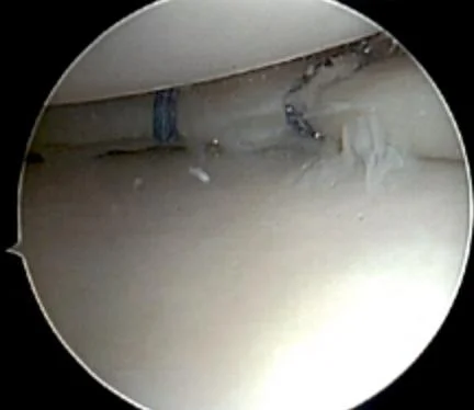
Meniscal repair surgery: step-by-step

Meniscal repair surgery is a minimally invasive procedure that aims to preserve the meniscus by stitching the torn edges back together. Unlike meniscectomy (where damaged tissue is removed), this technique is designed to maintain the natural function of the knee joint and reduce the long-term risk of arthritis.

Below is a general outline of the steps involved in arthroscopic meniscal repair:


1. Anaesthesia and patient preparation

The procedure is usually performed under general or spinal anaesthesia, depending on the patient’s health and preferences. The leg is positioned and sterilised, and a tourniquet may be applied to reduce bleeding and improve visibility inside the joint.

2. Arthroscopic assessment

Small incisions (keyholes) are made to insert an arthroscope (camera) and surgical instruments into the knee. The surgeon inspects the joint thoroughly, confirming:

  • The location, type, and severity of the meniscal tear
  • The condition of surrounding cartilage, ligaments (e.g. ACL), and joint surfaces

3. Preparing the tear for repair

The torn edges of the meniscus are gently debrided (cleaned) using a small shaver to promote healing. This allows better blood flow to the area and improves the chances of successful healing.

4. Suturing the tear

Dr Awwad uses specialised meniscal repair techniques depending on the tear pattern and location:

  • All-inside repair: Devices inserted entirely through the arthroscope place anchors and sutures internally. Often used for posterior horn tears.
  • Inside-out repair: Sutures are passed from inside the joint to outside the capsule, then tied under the skin. Suitable for mid-body or posterior tears.
  • Outside-in repair: Sutures are inserted from outside the joint into the tear. Often used for anterior horn tears.

Modern repair techniques use biodegradable implants or non-absorbable sutures designed to hold the meniscus securely while it heals.

5. Final inspection and closure

Once the repair is complete, Dr Awwad checks:

  • The stability of the repair
  • Range of motion to ensure there is no impingement
  • Bleeding control and irrigation of the joint

The instruments are removed, and the small incisions are closed with sutures or steri-strips. A sterile dressing is applied.

6. Post-operative care

Most patients go home on the same day or after an overnight stay. A rehabilitation program is initiated soon after surgery and tailored to the patient’s procedure. Recovery may take several months depending on the complexity of the repair and whether other procedures (e.g. ACL reconstruction) were performed at the same time.

Recovery and rehabilitation after meniscal repair

Recovery following a meniscal repair is carefully structured to allow the repaired tissue to heal properly while gradually restoring strength, mobility, and function. Rehabilitation will be tailored to your specific tear pattern and surgical details, but typically follows a staged protocol over several months.


Hospital stay and immediate post-operative period

  • Same-day surgery: Most patients go home the same day as their operation.
  • Bracing: A hinged knee brace is fitted immediately post-surgery to support the knee and protect the repair. The brace is worn locked in extension when walking and sleeping for the first 6 weeks.
  • Crutches: Crutches are required to assist with partial weight bearing for the first 6 weeks. The goal is to avoid excessive pressure on the healing meniscus.
  • Pain management: Medication and ice are used to control swelling and discomfort.

Weeks 0–6: Protection phase

  • Weight bearing: Partial weight bearing with crutches is allowed. The knee brace remains locked in full extension during walking.
  • Range of motion (ROM): Controlled passive and active-assisted ROM exercises are encouraged:
  • Knee flexion is gradually increased under physiotherapy supervision.
  • Aim for 0–90° of knee flexion by the end of week 6.
  • Muscle activation: Quadriceps and hamstring isometric exercises begin early to prevent atrophy.
  • Precautions: Avoid twisting, pivoting, or deep squats. No cycling or stair climbing unless advised by your physiotherapist.

Weeks 6–12: Transition phase

  • Discontinue brace: The hinged knee brace may be removed after 6 weeks, based on surgeon review.
  • Progressive loading: Transition to full weight bearing as tolerated. Crutches are weaned off gradually.
  • ROM goals: Increase knee flexion towards full range by 12 weeks.
  • Strengthening: Begin closed chain strengthening, balance, and proprioceptive exercises under physiotherapy guidance.

Months 3–6: Strengthening phase

  • Functional training: Progressive resistance training, single-leg balance, and neuromuscular control exercises are introduced.
  • Low-impact cardio: Stationary cycling, elliptical machines, and swimming are usually permitted.
  • Sport-specific drills: May begin light agility and sport-specific movement patterns around 4–5 months if recovery is progressing well.

Return to sport

  • Most patients return to non-contact sports and full activity between 5 to 6 months post-surgery, depending on:
  • Healing of the meniscus (confirmed via clinical assessment)
  • Absence of pain or swelling
  • Full strength and neuromuscular control
  • Contact or pivoting sports may be delayed longer to ensure graft integrity and joint readiness.

Physiotherapy support

  • Regular follow-up with a physiotherapist is critical.
  • Dr Awwad’s team will provide you with a structured protocol and liaise with your physio to ensure individualised progression.

Note: Timelines may vary based on your specific tear pattern (e.g. radial vs longitudinal) and surgical findings. Dr Awwad and the physiotherapist will guide your recovery and advise if any adjustments are needed to support optimal healing.

Benefits of repairing the meniscus

Whenever possible, repairing the meniscus instead of removing it may offer significant long-term advantages. A successful meniscal repair helps preserve the natural structure and function of your knee, supporting joint stability and long-term knee health.

Preservation of shock absorption

The meniscus acts as a cushion between the femur and tibia, absorbing the shock and load transmitted through the knee joint. By repairing the meniscus rather than removing it, this protective function is maintained, which may help reduce joint wear over time.

Support for long-term joint health

Studies have shown that preserving the meniscus may reduce the risk of developing osteoarthritis in the future. Repairing the meniscus allows the joint to function more naturally, potentially delaying or preventing degenerative changes.

Improved knee stability and biomechanics

The meniscus plays a critical role in guiding knee motion and providing stability, especially during twisting or pivoting movements. A repaired meniscus helps maintain normal joint mechanics and may lower the risk of further injury.

Better outcomes for younger or active patients

Meniscal repair is particularly beneficial for younger, athletic, or active individuals who place higher demands on their knees. Preserving the meniscus in this group may help support a return to sports and high-level activities with better long-term outcomes.

Minimally invasive technique

Meniscal repair is typically performed using arthroscopic (keyhole) surgery, which involves smaller incisions, less tissue disruption, and a lower risk of complications compared to open surgery. This can support a smoother recovery and faster return to function.

Note: Not all tears are suitable for repair. Dr Awwad will assess the size, location, and pattern of the tear to determine the most appropriate treatment approach.

Possible risks and complications of meniscal repair surgery

As with any surgical procedure, meniscal repair surgery carries some risks. While complications are uncommon, it’s important to be aware of them before proceeding.Possible risks may include:

Infection

Although sterile techniques are used, there is a small risk of infection in the joint or wound site.

These can occur in the leg after surgery. Preventative measures may be advised, particularly if other risk factors are present.

Scar tissue or inflammation can limit movement, particularly if rehabilitation is delayed.

Some discomfort is normal post-surgery, but ongoing pain may require further assessment.

Rarely, structures around the knee may be affected during surgery.

In some cases, the meniscus may not heal as expected, especially if the tear was complex, located in a poorly vascularised zone, or if post-op protocols are not followed.

This can happen due to reinjury, overuse, or non-compliance with rehabilitation.

Although repair aims to preserve joint health, some patients may develop osteoarthritis over time depending on their age, activity levels, and prior cartilage damage.

Although sterile techniques are used, there is a small risk of infection in the joint or wound site.These can occur in the leg after surgery. Preventative measures may be advised, particularly if other risk factors are present.
Scar tissue or inflammation can limit movement, particularly if rehabilitation is delayed.
Some discomfort is normal post-surgery, but ongoing pain may require further assessment.Rarely, structures around the knee may be affected during surgery.
In some cases, the meniscus may not heal as expected, especially if the tear was complex, located in a poorly vascularised zone, or if post-op protocols are not followed.
This can happen due to reinjury, overuse, or non-compliance with rehabilitation.
Although repair aims to preserve joint health, some patients may develop osteoarthritis over time depending on their age, activity levels, and prior cartilage damage.Dr Awwad will discuss these risks with you during the consent process. Following the recommended rehabilitation plan and attending scheduled follow-up appointments can help reduce the likelihood of complications and improve long-term outcomes.

FAQs about meniscal repair

What is the difference between a meniscal repair and a meniscectomy?

A meniscal repair involves stitching the torn meniscus back together to preserve as much of the tissue as possible. This approach aims to restore normal knee function and reduce the risk of arthritis later in life. A meniscectomy, by contrast, involves trimming and removing the damaged portion of the meniscus. Where possible, Dr Awwad aims to preserve and repair the meniscus rather than remove it.

Not all meniscus tears are suitable for repair. The decision depends on the location, pattern, and severity of the tear, as well as your age and activity level. Tears in the outer third of the meniscus (the “red zone”) have a better blood supply and are more likely to heal successfully after repair. During your consultation, Dr Awwad will review your imaging and provide personalised advice on whether repair is appropriate for you.

Pain is typically well controlled with medication after surgery. You may experience discomfort, swelling, and stiffness for a few days. Dr Awwad and the hospital team will provide a pain management plan and support your recovery with early physiotherapy and gradual return to movement.

Full recovery can take 3 to 6 months, depending on your age, overall health, and the specifics of the surgery. You will need to limit certain movements during the early recovery phase to allow the meniscus to heal. A structured rehabilitation program will be provided, and Dr Awwad will monitor your progress throughout.

You will usually be able to walk with the help of crutches in the first few weeks after surgery. Weight-bearing will be gradually increased under guidance from your physiotherapist and Dr Awwad. It is important to follow the prescribed movement restrictions to protect the repair.

Most patients return to sports between 5 to 6 months after surgery, depending on the type of activity and healing progress. High-impact and pivoting sports may require a longer recovery. Your physiotherapist and Dr Awwad will guide your return to activity.

Medicare covers a portion of the surgical fee. If you have private health insurance, some or all of your hospital and surgical costs may be covered, depending on your policy and level of cover. It’s best to confirm your rebates with your health fund ahead of surgery. Dr Awwad’s team can assist you with this.

In some cases, the tear may not heal completely, particularly if it was located in a region with poor blood supply. If symptoms persist, further imaging may be required, and in some cases, revision surgery or a partial meniscectomy might be considered.

Most patients return to sports between 5 to 6 months after surgery, depending on the type of activity and healing progress. High-impact and pivoting sports may require a longer recovery. Your physiotherapist and Dr Awwad will guide your return to activity.

Medicare covers a portion of the surgical fee. If you have private health insurance, some or all of your hospital and surgical costs may be covered, depending on your policy and level of cover. It’s best to confirm your rebates with your health fund ahead of surgery. Dr Awwad’s team can assist you with this.In some cases, the tear may not heal completely, particularly if it was located in a region with poor blood supply. If symptoms persist, further imaging may be required, and in some cases, revision surgery or a partial meniscectomy might be considered.
If you’re living with pain or stiffness that’s affecting your quality of life, a thorough assessment can help determine the cause and guide the right treatment path.

Book An Appointment