Understanding lateral hip weakness, pain, and instability related to gluteal muscle injuries
Hip abductor dysfunction refers to weakness, injury, or degeneration of the muscles responsible for stabilising the pelvis during walking and standing, particularly the gluteus medius and gluteus minimus. These muscles play a crucial role in maintaining balance and preventing the pelvis from tilting with each step. When the abductors are not functioning properly, patients may experience pain on the side of the hip, difficulty walking, limping (known as a Trendelenburg gait), or reduced stability.
This condition is commonly seen in middle aged to older adults, especially women, and may be caused by tendon tears, muscle atrophy, nerve irritation, or underlying joint conditions such as arthritis or hip dysplasia. In some cases, the dysfunction may mimic other causes of lateral hip pain, such as trochanteric bursitis, making accurate diagnosis essential.
Dr George Awwad provides a comprehensive assessment of hip abductor dysfunction and offers both non-surgical and surgical treatment pathways, depending on the underlying cause and severity of symptoms.
What are the hip abductor muscles and what do they do?
The hip abductor muscles are a group of muscles located on the outer portion of your hip and upper thigh. Their main role is to stabilise the pelvis and move the leg away from the body, a motion known as abduction. They are essential for maintaining balance while walking, standing on one leg, or climbing stairs.
Key Hip Abductor Muscles
1. Gluteus Medius
-
Located on the outer surface of the pelvis. -
The primary muscle responsible for hip abduction and stabilisation during walking. -
Prevents the pelvis from dropping on the opposite side when standing on one leg (Trendelenburg function).
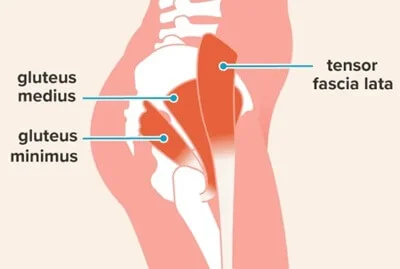
2. Gluteus Minimus
-
The smallest and deepest of the gluteal muscles, sitting beneath the gluteus medius. -
Assists in hip abduction and internal rotation. -
Works closely with the gluteus medius to maintain pelvic stability.
3. Tensor Fasciae Latae (TFL)
-
A small muscle on the outer thigh that assists with abduction and internal rotation of the hip. -
Connects to the iliotibial (IT) band, contributing to hip and knee movement.
Role in Pelvic Stability and Gait
-
These muscles are critical for pelvic alignment and balance during dynamic activities like walking, running, and single-leg standing. -
When functioning properly, the abductors ensure that the pelvis remains level, reducing strain on the lower back and knees. -
If these muscles are weak, torn, or not functioning correctly (as in hip abductor dysfunction), it can lead to a characteristic limping gait (Trendelenburg gait), pain on the outer hip, or instability.
Common causes of hip abductor dysfunction
Hip abductor dysfunction occurs when the muscles responsible for stabilising the hip, primarily the gluteus medius and gluteus minimus, become weak, torn, inflamed, or otherwise impaired. This condition can result in pain, limping, reduced mobility, and difficulty with daily activities such as walking or climbing stairs.
1. Tendon Tears or Degeneration
One of the most common causes is a partial or full-thickness tear of the gluteus medius or minimus tendons, often likened to a “rotator cuff tear of the hip.” These tears may result from:
-
Chronic overuse (e.g. repetitive walking or running) -
Age-related degeneration -
Previous trauma or falls -
Poor biomechanics or compensatory gait patterns
2. Tendinopathy or Inflammation
Chronic inflammation or degeneration of the tendons (gluteal tendinopathy) can lead to pain and weakness. This condition is often associated with:
-
Greater trochanteric pain syndrome (GTPS) -
Repetitive strain or overload -
Impaired healing response in older adults
3. Muscle Atrophy or Weakness
Generalised weakness or disuse of the abductor muscles can develop over time due to:
-
Sedentary lifestyle -
Hip osteoarthritis -
Previous surgery (e.g. hip replacement) -
Neurological conditions such as stroke or nerve injury
4. Nerve Impairment
Damage or compression of the superior gluteal nerve, which innervates the abductors, may result in muscle weakness and dysfunction. This may occur due to:
-
Hip or pelvic trauma -
Complications from surgery -
Tumours or mass effect in the pelvic region
5. Biomechanical and Structural Abnormalities
Conditions that alter the normal alignment or mechanics of the hip can increase the load on the abductors and predispose them to dysfunction. Examples include:
-
Hip dysplasia -
Leg length discrepancy -
Pelvic tilt or abnormal gait patterns -
Lumbar spine pathology affecting nerve function or posture
Signs and symptoms of abductor dysfunction
Hip abductor dysfunction can present with a range of symptoms, from subtle weakness to severe pain and mobility limitations. Because the abductor muscles play a crucial role in stabilising the pelvis during walking and standing, dysfunction can significantly impact your day-to-day activities.
1. Lateral hip pain
-
The most common complaint is pain on the outside of the hip, often over the greater trochanter (the bony prominence at the side of your hip). -
This pain may worsen:
-
With prolonged standing -
While lying on the affected side -
During activities such as climbing stairs or walking up hills
2. Limping or gait abnormalities
During your consultation, Dr Awwad will:
-
Weakness in the abductor muscles can lead to a Trendelenburg gait, where the pelvis drops on the opposite side during walking. -
Patients may unconsciously shift their torso over the affected side to compensate, creating a waddling or lurching motion.
3. Muscle weakness
-
You may notice difficulty standing on one leg (especially on the affected side), or a sense of instability when walking. -
Climbing stairs, getting up from a chair, or rising from a squat may become more challenging.
4. Tenderness and reduced range of motion
-
Pressing on the lateral aspect of the hip often elicits discomfort. -
Some individuals experience stiffness or reduced hip mobility, particularly when trying to move the leg outward or backward.
5. Referred pain
-
Pain may radiate down the outside of the thigh or into the buttock. -
It is important to differentiate this from other conditions such as sciatica or hip osteoarthritis.
How hip abductor dysfunction is diagnosed
Diagnosing hip abductor dysfunction involves a thorough clinical assessment, combined with targeted imaging if needed. Dr George Awwad will take into account your symptoms, functional limitations, medical history, and examination findings to accurately identify the underlying issue.
1. Clinical history
Your consultation will begin with a detailed discussion of your symptoms, including:
-
Your consultation will begin with a detailed discussion of your symptoms, including: -
Activities that aggravate or relieve the discomfort -
Duration of symptoms and any history of trauma or overuse -
Difficulty with daily activities such as walking, climbing stairs, or lying on the affected side
Understanding your lifestyle and activity level helps guide diagnosis and tailor management options.
2. Physical examination
During the examination, Dr Awwad will perform specific tests to assess the function of your abductor muscles and rule out other causes of lateral hip pain:
-
Trendelenburg test: Standing on one leg to check for pelvic drop, which indicates weakness in the gluteus medius. -
Palpation: Tenderness over the greater trochanter may suggest associated bursitis or tendon involvement. -
Strength testing: Manual resistance tests can reveal abductor weakness or pain. -
Gait assessment: Observing your walking pattern helps identify compensatory movements or limping.
3. Imaging tests
If the clinical findings suggest abductor dysfunction, imaging may be used to confirm the diagnosis or identify structural issues such as tendon tears:
-
X-rays: Help rule out underlying joint degeneration, fractures, or bony abnormalities. -
Ultrasound: Useful for assessing superficial tendon tears or bursitis. -
MRI: Provides detailed imaging of the hip tendons and muscles and is particularly helpful for detecting partial or full-thickness tears of the gluteus medius or minimus tendons.
4. Diagnostic injection (if needed)
In complex cases, a local anaesthetic and corticosteroid injection into the bursa may help distinguish between bursitis and true abductor tendon pathology. If pain relief is achieved, this may confirm that the pain source is related to bursal inflammation.
Non-surgical treatment options for hip abductor dysfunction
In many cases, hip abductor dysfunction can be managed without surgery, particularly when the condition is caught early or involves partial tendon injury, overuse, or muscular imbalance. The goal of non-surgical management is to reduce pain, restore function, and prevent further deterioration of the hip stabilisers.
1. Physiotherapy and targeted exercise
A structured physiotherapy program is often the cornerstone of treatment. Your physiotherapist may focus on:
-
Strengthening exercises for the gluteus medius and minimus -
Pelvic stability training to address compensation patterns -
Stretching exercises for surrounding muscles like the iliotibial band and hip flexors -
Gait re-education to reduce strain on weakened abductors
2. Activity modification
Reducing or temporarily avoiding aggravating activities may help control symptoms. This may include:
-
Limiting prolonged walking or standing -
Avoiding stairs or hills where possible -
Changing your sleeping position (e.g. avoiding lying on the affected side)
3. Anti-inflammatory medications
Non-steroidal anti-inflammatory drugs (NSAIDs), such as ibuprofen or meloxicam, may assist with pain and inflammation—particularly during flare-ups. These are typically used short-term and under the guidance of your GP or specialist.
4. Corticosteroid injections
If conservative measures do not provide sufficient relief, an ultrasound-guided corticosteroid injection may be considered. This can:
-
Reduce inflammation and pain in the surrounding bursa or tendon sheath -
Improve comfort to allow progression with physiotherapy -
Assist in confirming the pain source
Injections may be repeated cautiously, but overuse can weaken the tendon, so this is not typically a long-term strategy.
5. Shockwave therapy
Extracorporeal shockwave therapy (ESWT) may be recommended in some cases, particularly if tendon thickening or partial tearing is present. This non-invasive treatment involves pulsed acoustic waves aimed at:
-
Stimulating blood flow -
Promoting healing of the tendon tissue -
Reducing chronic pain
Evidence for ESWT in hip abductor tendinopathy is growing, though it is generally reserved for persistent symptoms.
Surgical options for persistent or severe cases
When non-surgical treatments do not sufficiently relieve pain or restore function and imaging confirms significant damage or tearing of the hip abductor tendons, surgical intervention may be considered. Surgery is usually reserved for patients with full-thickness tendon tears, persistent instability, or failed conservative management over several months.
1. Hip abductor tendon repair
This is the most common surgical procedure performed for significant abductor dysfunction.
Key steps include:
-
Reattaching the torn gluteus medius and/or minimus tendon to the greater trochanter (outer part of the hip bone) -
Removing any damaged or degenerated tendon tissue -
Anchoring the tendon using sutures or bone anchors
This procedure may be performed as:
-
Open surgery for direct visualisation of the tear -
Minimally invasive (endoscopic or arthroscopic) surgery, where small incisions and a camera are used, potentially reducing recovery time
2. Trochanteric bursectomy
Often performed alongside tendon repair, this procedure involves removing the inflamed trochanteric bursa. This may help reduce ongoing pain and irritation from surrounding tissue and scar formation.
3. Tendon grafting or augmentation
In select cases, biological or synthetic grafts may be used to:
-
Reinforce the repair -
Bridge tendon defects that cannot be fully closed -
Provide a scaffold to support tendon healing
This may be considered for patients with poor-quality tissue or revision surgeries.
1. Hip abductor tendon repair
This is the most common surgical procedure performed for significant abductor dysfunction.Key steps include:
-
Reattaching the torn gluteus medius and/or minimus tendon to the greater trochanter (outer part of the hip bone) -
Removing any damaged or degenerated tendon tissue -
Anchoring the tendon using sutures or bone anchors
This procedure may be performed as:
-
Open surgery for direct visualisation of the tear -
Minimally invasive (endoscopic or arthroscopic) surgery, where small incisions and a camera are used, potentially reducing recovery time
2. Trochanteric bursectomy
Often performed alongside tendon repair, this procedure involves removing the inflamed trochanteric bursa. This may help reduce ongoing pain and irritation from surrounding tissue and scar formation.
3. Tendon grafting or augmentation
In select cases, biological or synthetic grafts may be used to:
-
Reinforce the repair -
Bridge tendon defects that cannot be fully closed -
Provide a scaffold to support tendon healing
This may be considered for patients with poor-quality tissue or revision surgeries.
Frequently asked questions about hip abductor dysfunction
What are the hip abductor muscles, and why are they important?
The hip abductor muscles, primarily the gluteus medius and gluteus minimus are responsible for stabilising the pelvis and allowing side-to-side leg movement. They play a critical role in walking, climbing stairs, and maintaining balance.
What causes hip abductor dysfunction?
Common causes include tendon tears, gluteal tendinopathy, trauma, post-operative changes after hip replacement, nerve injury (particularly involving the superior gluteal nerve), and overuse in athletes or active individuals.
How do I know if I have a hip abductor issue?
You may experience pain on the outside of the hip, a limp when walking, difficulty balancing on one leg, or a noticeable drop in the pelvis on the opposite side during walking (positive Trendelenburg sign). In some cases, patients report weakness when climbing stairs or rising from a chair.
What imaging is used to diagnose hip abductor dysfunction?
Dr Awwad may recommend imaging such as MRI or ultrasound to assess the gluteal tendons, surrounding soft tissues, and any signs of inflammation or tearing. These scans provide a detailed view of tendon quality and extent of damage.
Will I need surgery?
Not always. Many cases respond well to physiotherapy, anti-inflammatory medication, or corticosteroid injections. If non-surgical treatment fails or there is a complete tear of the gluteal tendons, surgical repair may be considered.
The hip abductor muscles, primarily the gluteus medius and gluteus minimus are responsible for stabilising the pelvis and allowing side-to-side leg movement. They play a critical role in walking, climbing stairs, and maintaining balance.
Common causes include tendon tears, gluteal tendinopathy, trauma, post-operative changes after hip replacement, nerve injury (particularly involving the superior gluteal nerve), and overuse in athletes or active individuals.
You may experience pain on the outside of the hip, a limp when walking, difficulty balancing on one leg, or a noticeable drop in the pelvis on the opposite side during walking (positive Trendelenburg sign). In some cases, patients report weakness when climbing stairs or rising from a chair.Dr Awwad may recommend imaging such as MRI or ultrasound to assess the gluteal tendons, surrounding soft tissues, and any signs of inflammation or tearing. These scans provide a detailed view of tendon quality and extent of damage.
Not always. Many cases respond well to physiotherapy, anti-inflammatory medication, or corticosteroid injections. If non-surgical treatment fails or there is a complete tear of the gluteal tendons, surgical repair may be considered.
What does surgery for hip abductor dysfunction involve?
Surgical repair typically involves reattaching the torn tendons to the greater trochanter using sutures and anchors. In some cases, tissue grafts may be needed. Dr Awwad will discuss the appropriate technique based on your condition and goals.
What is the recovery like after surgery?
Recovery can involve using crutches for several weeks, followed by a gradual rehabilitation program guided by a physiotherapist. Full recovery and return to activity may take several months, depending on the severity of the injury and your overall health.
Can hip abductor dysfunction come back after treatment?
With appropriate management and adherence to a rehabilitation plan, most patients experience long-term improvement. However, re-injury can occur, particularly if underlying risk factors (e.g., muscle imbalance or poor biomechanics) are not addressed.
Is this condition common after hip replacement?
Yes, abductor dysfunction can occur after hip replacement, particularly with lateral or posterior approaches. Dr Awwad assesses each case carefully to determine whether the dysfunction is due to tendon detachment, muscle damage, or nerve involvement.
When should I seek specialist review?
If you are experiencing persistent outer hip pain, weakness, or difficulty walking that isn’t improving with rest or basic physiotherapy, it may be time to book a consultation. Early assessment may help prevent further deterioration and support a better recovery outcome.
Surgical repair typically involves reattaching the torn tendons to the greater trochanter using sutures and anchors. In some cases, tissue grafts may be needed. Dr Awwad will discuss the appropriate technique based on your condition and goals.
Recovery can involve using crutches for several weeks, followed by a gradual rehabilitation program guided by a physiotherapist. Full recovery and return to activity may take several months, depending on the severity of the injury and your overall health.
With appropriate management and adherence to a rehabilitation plan, most patients experience long-term improvement. However, re-injury can occur, particularly if underlying risk factors (e.g., muscle imbalance or poor biomechanics) are not addressed.Yes, abductor dysfunction can occur after hip replacement, particularly with lateral or posterior approaches. Dr Awwad assesses each case carefully to determine whether the dysfunction is due to tendon detachment, muscle damage, or nerve involvement.If you are experiencing persistent outer hip pain, weakness, or difficulty walking that isn’t improving with rest or basic physiotherapy, it may be time to book a consultation. Early assessment may help prevent further deterioration and support a better recovery outcome. If you’re living with hip pain or stiffness that’s affecting your quality of life, a thorough assessment can help determine the cause and guide the right treatment path.
Book An Appointment