Surgical treatment for fractures of the knee joint
Knee fractures are serious injuries that involve a break in one or more of the bones that make up the knee joint, most commonly the patella (kneecap), distal femur (thighbone), or proximal tibia (shinbone). These injuries are often the result of high-impact trauma, falls, or sporting accidents, and may affect not only the bone but also nearby cartilage, ligaments, and soft tissues.
Knee fracture surgery may be recommended to restore joint stability, preserve function, and support long-term mobility. Treatment typically involves realigning the broken bone fragments and securing them with internal fixation devices such as plates, screws, or rods. Early and accurate surgical intervention aims to reduce the risk of complications such as stiffness, arthritis, or prolonged disability.
On this page, you’ll find detailed information about the types of knee fractures, when surgery may be required, the procedures involved, and what to expect during recovery. Dr George Awwad will guide you through each step of your care, tailoring your treatment to your individual injury and functional goals.
What are the different types of knee fractures?
Your knee is a complex joint made up of bones, cartilage, ligaments, and tendons that work together to provide stability and movement. A fracture in any of the bones forming the knee can significantly impact your ability to walk, bear weight, or move comfortably. The type of knee fracture you experience will influence your treatment plan and recovery process.
Below is an overview of the most common types of knee fractures:
Patella (kneecap) fractures
The patella is a small, triangular bone that sits at the front of your knee. It protects the joint and helps your quadriceps muscle extend the leg. A patellar fracture usually results from a direct blow to the knee such as a fall or collision and may involve a clean break or a shattered bone. These fractures can be displaced (the bone fragments are separated) or non-displaced (the bone remains aligned).
Distal femur fractures (lower end of the thigh bone)
The distal femur is the lower part of the thigh bone that connects to the top of the knee joint. Fractures in this area often occur from high-energy trauma (like motor vehicle accidents) or low-energy injuries in older adults with osteoporosis. These fractures can extend into the knee joint, potentially damaging cartilage and causing long-term joint issues if not treated properly.
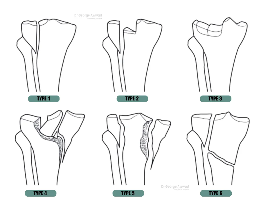
Proximal tibia fractures (upper end of the shin bone)
The proximal tibia forms the lower part of the knee joint and supports much of your body weight. Fractures here are sometimes called tibial plateau fractures and can affect the stability and alignment of the knee. These injuries are often seen in sports injuries or falls and may be associated with ligament or meniscal damage due to the force required to fracture this strong bone.
Tibial spine fractures
The tibial spine is a bony ridge where the anterior cruciate ligament (ACL) attaches to the tibia. These fractures are more common in children and adolescents and may mimic ACL injuries. Depending on the degree of displacement, they may require surgical fixation to restore joint stability and prevent long-term problems.
Segond fractures
Although less common, a Segond fracture is a small avulsion (pull-off) fracture of the lateral tibial plateau, typically associated with ACL injuries. It is considered a marker for significant internal knee trauma and may indicate the need for further surgical intervention.
Multiple and complex knee fractures
Some patients may experience fractures involving more than one bone in the knee, or multiple fracture lines within the same bone. These are often classified as complex or comminuted fractures and require a tailored surgical approach to restore joint anatomy and function.
Understanding your specific type of fracture is the first step in developing a treatment plan tailored to your injury. During your consultation, Dr George Awwad will review your imaging and explain the type of fracture you have, how it affects your joint, and which treatment option is most appropriate for your recovery.
Causes and risk factors for knee fractures
Knee fractures often occur when the bones that form the knee joint such as the femur, tibia, or patella are exposed to excessive force or trauma. These injuries can range from simple cracks to complex breaks that disrupt joint stability and function. Understanding the common causes and risk factors can help patients recognise how these injuries occur and what may increase their likelihood.
Multiple and complex knee fractures
1. High-impact trauma
Road traffic accidents, falls from height, and high-speed sports collisions are among the most frequent causes of knee fractures. These high-energy events can cause multiple or complex fractures, especially in younger individuals.
2. Preoperative Planning
Contact sports such as football, rugby, or skiing can place the knee at risk of traumatic fractures, particularly tibial plateau fractures and patellar fractures, often accompanied by ligament or cartilage injuries.
3. Falls and slips
In older adults, a simple fall, particularly onto a hard surface, can result in a knee fracture due to reduced bone density and balance issues.
4. Twisting or rotational force
Sudden pivoting movements or an awkward landing can cause fractures, particularly if the bones are already compromised. This is sometimes seen in conjunction with ligament tears.
5. Direct impact or blunt force trauma
A direct blow to the knee such as from a fall onto a hard surface or being struck by an object can fracture the patella or other knee bones.
Road traffic accidents, falls from height, and high-speed sports collisions are among the most frequent causes of knee fractures. These high-energy events can cause multiple or complex fractures, especially in younger individuals.Contact sports such as football, rugby, or skiing can place the knee at risk of traumatic fractures, particularly tibial plateau fractures and patellar fractures, often accompanied by ligament or cartilage injuries.In older adults, a simple fall, particularly onto a hard surface, can result in a knee fracture due to reduced bone density and balance issues.Sudden pivoting movements or an awkward landing can cause fractures, particularly if the bones are already compromised. This is sometimes seen in conjunction with ligament tears.
A direct blow to the knee such as from a fall onto a hard surface or being struck by an object can fracture the patella or other knee bones.
Risk factors that increase the likelihood of a knee fracture
1. Osteoporosis and low bone density
Weakened bones are more susceptible to fractures, even with lower-impact trauma. Older adults, particularly post-menopausal women, are at increased risk due to bone thinning.
2. Age
As people age, bone strength, balance, and muscle tone tend to decline, making knee fractures from falls more common.
3. Participation in high-risk activities
Individuals who engage in contact sports, extreme sports, or physically demanding work are more likely to sustain traumatic knee injuries.
4. Previous knee injuries
A history of knee injuries, especially if they were not properly treated or rehabilitated, can weaken the bone structure or alignment, increasing the chance of a future fracture.
5. Certain medical conditions
Conditions such as arthritis, cancer that spreads to the bone, or genetic bone disorders may predispose patients to fractures under otherwise minor stress.
Weakened bones are more susceptible to fractures, even with lower-impact trauma. Older adults, particularly post-menopausal women, are at increased risk due to bone thinning.As people age, bone strength, balance, and muscle tone tend to decline, making knee fractures from falls more common.
Individuals who engage in contact sports, extreme sports, or physically demanding work are more likely to sustain traumatic knee injuries.
A history of knee injuries, especially if they were not properly treated or rehabilitated, can weaken the bone structure or alignment, increasing the chance of a future fracture.
Conditions such as arthritis, cancer that spreads to the bone, or genetic bone disorders may predispose patients to fractures under otherwise minor stress.
If you suspect a knee fracture, early medical assessment and imaging are essential to determine the severity of the injury and guide appropriate treatment. Dr George Awwad will provide a thorough clinical review and recommend a personalised treatment plan to support your recovery and long-term joint health.
Symptoms of a fractured knee
A knee fracture may cause a range of symptoms depending on the severity, location, and number of bones affected. Prompt recognition of symptoms is important to help avoid further damage and begin appropriate treatment.
Pain and tenderness
Most people with a knee fracture experience sudden, intense pain at the time of injury. The pain is typically localised to the area of the fracture, such as around the kneecap, on the inner or outer side of the knee, or below the kneecap and is often worsened with any attempt to move, stand, or bear weight.
Swelling and bruising
Swelling is a common symptom and may develop quickly after the injury. This may be accompanied by visible bruising or discolouration around the knee joint and sometimes down the leg due to gravity.
Inability to bear weight
Depending on the type of fracture, it may be difficult or impossible to stand, walk, or put weight on the affected leg. In some cases, people may feel the knee ‘give way’ or feel unstable.
Reduced range of motion
You may find it hard or painful to bend or straighten the knee fully. This stiffness or loss of motion is often due to swelling, pain, or joint misalignment caused by the fracture.
Visible deformity
In more severe fractures, the knee may appear visibly deformed or out of alignment. This can include a sunken appearance of the kneecap (in patella fractures) or a change in the contour of the leg.
Grinding or clicking sensation
Some people report a grinding, popping, or clicking sensation in the knee after the injury. This may suggest damage to cartilage or joint surfaces in addition to the fracture.
Numbness or tingling
Although less common, some fractures may irritate or injure surrounding nerves or blood vessels, leading to numbness, tingling, or coolness in the lower leg or foot. These symptoms should be assessed urgently.
How are knee fractures diagnosed?
Accurate diagnosis of a knee fracture is essential to guide treatment and support the best possible recovery. Dr George Awwad uses a combination of clinical evaluation and imaging studies to assess the type, severity, and location of the fracture, along with any associated injuries to ligaments, cartilage, or tendons.
Medical history and clinical examination
Your consultation with Dr Awwad will begin with a detailed discussion about how the injury occurred. Information such as the mechanism of trauma (e.g. fall, motor vehicle accident, sports injury), your symptoms, and whether you were able to walk or bear weight afterward can provide important diagnostic clues.
Dr Awwad will then perform a physical examination to assess:
-
Local swelling, bruising, or deformity -
Tenderness over the bones or joint -
Range of motion and joint stability -
Circulation, sensation, and muscle strength in the leg and foot
If an open wound is present, the possibility of an open (compound) fracture will be assessed urgently.
X-rays (Radiographs)
Standard X-rays are usually the first step in confirming the presence of a fracture. They help visualise the bone structure, alignment, and whether the break is:
-
Displaced or non-displaced -
Simple or comminuted (multiple fragments) -
Intra-articular (extending into the knee joint)
Multiple views may be taken to ensure a full understanding of the fracture pattern, such as front-to-back (AP view), side-on (lateral view), and angled views if needed.
CT scan (Computed Tomography)
For more complex or joint-involving fractures (such as tibial plateau fractures), Dr Awwad may request a CT scan. This provides highly detailed cross-sectional images and 3D reconstructions to plan surgical repair more precisely.
MRI (Magnetic Resonance Imaging)
An MRI may be used when soft tissue injury is suspected in addition to the bone fracture. This may include damage to ligaments (ACL, PCL), menisci, or cartilage. It is especially helpful in high-impact injuries or when symptoms are not explained by X-ray findings alone.
Additional tests if required
In select cases, other tests such as vascular imaging (to assess blood flow) or bone scans (to detect stress fractures or hidden injuries) may be considered.
Medical history and clinical examination
Your consultation with Dr Awwad will begin with a detailed discussion about how the injury occurred. Information such as the mechanism of trauma (e.g. fall, motor vehicle accident, sports injury), your symptoms, and whether you were able to walk or bear weight afterward can provide important diagnostic clues.Dr Awwad will then perform a physical examination to assess:
-
Local swelling, bruising, or deformity -
Tenderness over the bones or joint -
Range of motion and joint stability -
Circulation, sensation, and muscle strength in the leg and foot
If an open wound is present, the possibility of an open (compound) fracture will be assessed urgently.
X-rays (Radiographs)
Standard X-rays are usually the first step in confirming the presence of a fracture. They help visualise the bone structure, alignment, and whether the break is:
-
Displaced or non-displaced -
Simple or comminuted (multiple fragments) -
Intra-articular (extending into the knee joint)
Multiple views may be taken to ensure a full understanding of the fracture pattern, such as front-to-back (AP view), side-on (lateral view), and angled views if needed.
CT scan (Computed Tomography)
For more complex or joint-involving fractures (such as tibial plateau fractures), Dr Awwad may request a CT scan. This provides highly detailed cross-sectional images and 3D reconstructions to plan surgical repair more precisely.
MRI (Magnetic Resonance Imaging)
An MRI may be used when soft tissue injury is suspected in addition to the bone fracture. This may include damage to ligaments (ACL, PCL), menisci, or cartilage. It is especially helpful in high-impact injuries or when symptoms are not explained by X-ray findings alone.
Additional tests if required
In select cases, other tests such as vascular imaging (to assess blood flow) or bone scans (to detect stress fractures or hidden injuries) may be considered.
When is surgery typically recommended for a knee fracture?
Surgery is typically recommended for knee fractures when the broken bone is displaced, unstable, or involves the joint surface in a way that may affect long-term knee function. Dr George Awwad carefully assesses the type of fracture, your overall health, activity level, and the risk of complications before recommending surgical treatment.
Common reasons for surgical intervention
Surgery may be advised in the following situations:
-
Displaced fractures: If the bone fragments have moved out of alignment, surgical fixation is often needed to restore the correct anatomy. -
Intra-articular fractures: When the break extends into the knee joint (such as a tibial plateau fracture), surgery helps ensure joint congruency to reduce the risk of arthritis. -
Unstable fractures: Some fractures are inherently unstable due to muscle and ligament forces pulling on the bone, making non-operative treatment difficult. -
Multiple fragments (comminuted fractures): Surgery can help reassemble and stabilise complex fracture patterns. -
Open fractures: If the bone has broken through the skin, urgent surgery is required to clean the wound and fix the fracture to reduce infection risk. -
Failure of non-operative management: If a fracture does not heal as expected with bracing or casting, surgery may become necessary.
Surgical goals
The main goals of surgery are to:
-
Realign the fractured bones (known as reduction) -
Secure them with implants such as plates, screws, or rods -
Restore the joint surface, if involved -
Support early movement to reduce stiffness and promote recovery
Dr Awwad will discuss whether surgical treatment is likely to improve your outcome based on your specific injury and circumstances. In all cases, the decision is made with careful consideration of the potential benefits and risks, tailored to your needs.
Types of surgical procedures used to treat knee fractures
The type of surgery used to treat a knee fracture depends on the location, severity, and pattern of the fracture, as well as the patient’s age, bone quality, and activity level. Dr George Awwad uses a range of evidence-based surgical techniques designed to restore alignment, stabilise the fracture, and support early rehabilitation.
Open reduction and internal fixation (ORIF)
Road traffic accidents, falls from height, and high-speed sports collisions are among the most frequent causes of knee fractures. These high-energy events can cause multiple or complex fractures, especially in younger individuals.
Intramedullary nailing
Contact sports such as football, rugby, or skiing can place the knee at risk of traumatic fractures, particularly tibial plateau fractures and patellar fractures, often accompanied by ligament or cartilage injuries.
Patella tension band wiring or screw fixation
In older adults, a simple fall, particularly onto a hard surface, can result in a knee fracture due to reduced bone density and balance issues.
External fixation
Sudden pivoting movements or an awkward landing can cause fractures, particularly if the bones are already compromised. This is sometimes seen in conjunction with ligament tears.
Arthroscopically assisted fixation
A direct blow to the knee such as from a fall onto a hard surface or being struck by an object can fracture the patella or other knee bones.
Road traffic accidents, falls from height, and high-speed sports collisions are among the most frequent causes of knee fractures. These high-energy events can cause multiple or complex fractures, especially in younger individuals.Contact sports such as football, rugby, or skiing can place the knee at risk of traumatic fractures, particularly tibial plateau fractures and patellar fractures, often accompanied by ligament or cartilage injuries.In older adults, a simple fall, particularly onto a hard surface, can result in a knee fracture due to reduced bone density and balance issues.Sudden pivoting movements or an awkward landing can cause fractures, particularly if the bones are already compromised. This is sometimes seen in conjunction with ligament tears.
A direct blow to the knee such as from a fall onto a hard surface or being struck by an object can fracture the patella or other knee bones.Each surgical approach is tailored to the patient’s specific injury and goals. Dr Awwad will explain the rationale behind the chosen method and what to expect during your consultation.
What happens during knee fracture surgical procedure?
Surgery for knee fractures is tailored to the type and severity of the injury. The aim is to realign and stabilise the fractured bones, protect the joint surface, and restore normal knee function. Dr George Awwad uses modern techniques and implants to support accurate reconstruction and long-term outcomes.
Before surgery
-
Imaging: X-rays and often a CT scan are used to understand the exact fracture pattern. -
Pre-operative planning: Dr Awwad will create a surgical plan based on your injury, overall health, and activity level. -
Anaesthesia: The procedure is typically performed under general anaesthetic or regional (spinal) anaesthesia.
Advanced, multi-compartment arthritis
Osteotomy is most effective when only one compartment of the knee is affected by arthritis. If the damage extends to multiple areas of the joint, a realignment procedure is unlikely to provide sufficient long-term relief and may even worsen symptoms.
Severe stiffness or limited range of motion
Patients with significantly reduced knee movement or longstanding stiffness may not achieve a good outcome with osteotomy, as the surgery is less effective when joint mobility is already compromised.
Inflammatory arthritis or systemic joint disease
Conditions like rheumatoid arthritis or other inflammatory joint diseases often affect the entire knee, making joint realignment procedures less suitable. These patients may benefit more from joint replacement surgery or other medical therapies.
Poor bone quality
Adequate bone strength is necessary for the osteotomy to heal effectively. People with osteoporosis or other conditions that weaken bone may be at increased risk of non-union or implant failure.
Unrealistic expectations
Patients who expect a complete return to high-impact sports or full joint restoration may be disappointed. Osteotomy is designed to delay knee replacement and improve quality of life, not to “cure” arthritis or create a perfect joint.
After surgery
-
You will be monitored in the recovery unit before returning to the ward. -
Pain management and early movement strategies will begin under the guidance of Dr Awwad and the physiotherapy team. -
You may be advised to avoid putting weight on the leg for a period, depending on the stability of the repair and the healing progress.
What is the recovery process like after knee fracture surgery
Recovery from knee fracture surgery is a gradual process that involves rest, bone healing, physiotherapy, and a structured return to movement and function. The exact timeline and rehabilitation plan can vary depending on the type of fracture, the surgical approach, your age, and your general health.
Hospital stay and immediate post-operative care
-
Most patients stay in hospital for 1 to 3 days, depending on the complexity of the surgery and initial mobility. -
Pain and swelling are managed with prescribed medications and supportive measures like elevation and ice. -
A brace, splint, or cast may be used to protect the knee and limit motion while the bone heals.
Weight-bearing and mobility
-
Whether you can put weight on your leg immediately after surgery depends on the type and stability of the fracture. -
Dr Awwad will provide specific guidance—some patients may need to remain non-weight-bearing for several weeks, while others may begin partial or full weight-bearing earlier. -
Crutches or a walking frame are often used initially to support mobility and safety.
Physiotherapy and rehabilitation
-
A physiotherapy program typically begins during your hospital stay and continues after discharge. -
Early exercises may focus on:
-
Maintaining range of motion in the hip, ankle, and unaffected joints -
Gentle knee mobilisation (as permitted) -
Muscle activation and strengthening
-
Over time, the program will progress to:
-
Weight-bearing exercises -
Balance and coordination retraining -
Functional activities such as walking, climbing stairs, and returning to sport (if applicable)
Bone healing and imaging follow-up
-
X-rays are taken periodically to monitor how well the fracture is healing. -
It can take around 6 to 12 weeks for the bone to unite, although this varies based on the fracture type and patient factors. -
Full recovery may take several months, especially in more complex or intra-articular fractures.
Returning to daily activities
-
Driving: You may return to driving once you can fully bear weight, safely control the pedals, and are no longer taking strong pain medication. This is typically around 6–8 weeks but may vary. -
Work: Office-based jobs may be resumed in 2–6 weeks, while physically demanding roles may require 3 months or longer. -
Sport: High-impact activities and sports are usually delayed for at least 4–6 months to reduce the risk of re-injury.
Ongoing care with Dr Awwad
Dr Awwad will review your progress at regular intervals and adjust your recovery plan as needed. Long-term follow-up may be required for more complex fractures to monitor joint health and function.
Potential risks or complications of knee fracture surgery
Like all surgical procedures, knee fracture surgery carries some risks. Dr George Awwad takes every precaution to reduce the likelihood of complications and will provide personalised information based on your overall health, the type of fracture, and the surgical technique used.
1. Infection
-
Post-operative infection may occur at the skin incision or around the implanted hardware. -
This is generally minimised through sterile techniques and may be treated with antibiotics if detected early. -
In rare cases, a deep infection may require additional surgery.
2. Blood clots (Deep Vein Thrombosis or Pulmonary Embolism)
-
Surgery and reduced mobility increase the risk of blood clots forming in the leg veins. -
Preventive measures may include blood-thinning medication, compression stockings, and early mobilisation.
3. Delayed bone healing or nonunion
-
Some fractures may take longer than expected to heal, particularly in patients who smoke or have certain medical conditions. -
In rare cases, the bone may not heal at all (nonunion), which could require further surgical intervention.
4. Malunion
-
This occurs when the fracture heals in an incorrect position, which may affect joint alignment or function. -
In some cases, revision surgery may be needed to restore optimal alignment.
5. Implant-related issues
-
Plates, screws, or other fixation devices are commonly used to stabilise the fracture. -
These implants can sometimes cause irritation, loosening, or may need removal after the bone has healed.
6. Joint stiffness or reduced range of motion
-
Scar tissue or prolonged immobilisation can result in knee stiffness. -
Physiotherapy is important to help restore joint mobility and minimise long-term stiffness.
7. Post-traumatic arthritis
-
Damage to the cartilage or joint surface during the fracture can increase the risk of developing osteoarthritis in the future. -
In some cases, joint replacement surgery may be needed later in life.
8. Nerve or blood vessel injury
-
Although rare, surrounding nerves or blood vessels may be injured during the fracture or surgery, which can cause numbness, weakness, or circulation issues.
9. Persistent pain or swelling
-
Some patients may experience ongoing discomfort, even after the fracture has healed, especially if there is residual soft tissue damage or early arthritis.
10. Anaesthesia-related risks
-
These include rare but possible complications such as allergic reactions or respiratory issues, which are managed by your anaesthetic team.
-
Post-operative infection may occur at the skin incision or around the implanted hardware. -
This is generally minimised through sterile techniques and may be treated with antibiotics if detected early. -
In rare cases, a deep infection may require additional surgery.
-
Surgery and reduced mobility increase the risk of blood clots forming in the leg veins. -
Preventive measures may include blood-thinning medication, compression stockings, and early mobilisation.
-
Some fractures may take longer than expected to heal, particularly in patients who smoke or have certain medical conditions. -
In rare cases, the bone may not heal at all (nonunion), which could require further surgical intervention.
-
This occurs when the fracture heals in an incorrect position, which may affect joint alignment or function. -
In some cases, revision surgery may be needed to restore optimal alignment.
-
Plates, screws, or other fixation devices are commonly used to stabilise the fracture. -
These implants can sometimes cause irritation, loosening, or may need removal after the bone has healed.
-
Scar tissue or prolonged immobilisation can result in knee stiffness. -
Physiotherapy is important to help restore joint mobility and minimise long-term stiffness.
-
Damage to the cartilage or joint surface during the fracture can increase the risk of developing osteoarthritis in the future. -
In some cases, joint replacement surgery may be needed later in life.
-
Although rare, surrounding nerves or blood vessels may be injured during the fracture or surgery, which can cause numbness, weakness, or circulation issues.
-
Some patients may experience ongoing discomfort, even after the fracture has healed, especially if there is residual soft tissue damage or early arthritis.
-
These include rare but possible complications such as allergic reactions or respiratory issues, which are managed by your anaesthetic team.
Knee Fracture Surgery FAQs
Below are answers to common questions patients often ask when considering or recovering from knee fracture surgery. These FAQs are designed to support your understanding and help you feel more informed throughout your treatment journey with Dr George Awwad.
Can I go private for my knee fracture surgery?
Yes, you may be able to undergo knee fracture surgery in the private hospital system. This generally requires a valid referral from your GP or emergency department and depends on the availability of a surgeon with admitting rights. Going private may allow for quicker access to surgery, a choice of treating surgeon, and potentially shorter hospital stays. Dr George Awwad operates in both public and private settings, and his team can guide you through the referral and admission process.
How long does it take to recover from knee fracture surgery?
Recovery time varies depending on the type and severity of the fracture, the surgical method used, and your general health. In most cases, initial healing takes 6 to 12 weeks, but full recovery, including return to sport or high-impact activities may take several months. Dr Awwad will tailor your post-operative plan and provide a detailed rehabilitation protocol to support optimal healing.
Will I need physiotherapy after surgery?
Yes, physiotherapy plays a critical role in restoring movement, strength, and confidence in your knee after fracture surgery. A physiotherapy program will typically begin with gentle range-of-motion exercises and progress to strengthening and functional training over time.
Can metal plates or screws stay in the body permanently?
In many cases, surgical implants such as plates, screws, or rods can remain in the body long-term without causing issues. However, if they become painful, restrict movement, or interfere with nearby tissues, removal may be considered after the bone has fully healed.
When can I return to work or driving after surgery?
This depends on the type of surgery, your occupation, and whether your driving leg is affected.
-
Office-based work: often possible within 2 to 6 weeks. -
Manual or physical jobs: may require 8 to 12 weeks or longer. -
Driving: typically allowed once you are no longer using crutches and can safely control the vehicle — usually after 6 to 8 weeks. Dr Awwad will provide individual guidance based on your recovery.
Yes, you may be able to undergo knee fracture surgery in the private hospital system. This generally requires a valid referral from your GP or emergency department and depends on the availability of a surgeon with admitting rights. Going private may allow for quicker access to surgery, a choice of treating surgeon, and potentially shorter hospital stays. Dr George Awwad operates in both public and private settings, and his team can guide you through the referral and admission process.
Recovery time varies depending on the type and severity of the fracture, the surgical method used, and your general health. In most cases, initial healing takes 6 to 12 weeks, but full recovery, including return to sport or high-impact activities may take several months. Dr Awwad will tailor your post-operative plan and provide a detailed rehabilitation protocol to support optimal healing.
Yes, physiotherapy plays a critical role in restoring movement, strength, and confidence in your knee after fracture surgery. A physiotherapy program will typically begin with gentle range-of-motion exercises and progress to strengthening and functional training over time.
In many cases, surgical implants such as plates, screws, or rods can remain in the body long-term without causing issues. However, if they become painful, restrict movement, or interfere with nearby tissues, removal may be considered after the bone has fully healed.
This depends on the type of surgery, your occupation, and whether your driving leg is affected.
-
Office-based work: often possible within 2 to 6 weeks. -
Manual or physical jobs: may require 8 to 12 weeks or longer. -
Driving: typically allowed once you are no longer using crutches and can safely control the vehicle — usually after 6 to 8 weeks. Dr Awwad will provide individual guidance based on your recovery.
Is knee fracture surgery painful?
You may experience discomfort following surgery, particularly in the first few days. Dr Awwad and his anaesthetic team will provide a tailored pain management plan that may include oral medication, local anaesthetic blocks, and strategies for reducing swelling and stiffness.
What happens if the bone doesn’t heal properly?
In rare cases, a bone may not heal completely (nonunion) or may heal in a less-than-optimal position (malunion). If this causes pain or functional problems, additional surgery such as revision fixation or corrective osteotomy may be needed.
Can knee fracture surgery prevent future arthritis?
Accurate fracture reduction and joint surface restoration aim to reduce the risk of developing post-traumatic arthritis. However, any injury involving cartilage damage may increase your risk of arthritis over time, regardless of surgical treatment.
How will I know when the bone is fully healed?
Healing is assessed through clinical review and repeat imaging (usually X-rays). Signs of healing include pain reduction, improved function, and radiological evidence of bone consolidation. Dr Awwad will monitor your progress closely during follow-up visits.
You may experience discomfort following surgery, particularly in the first few days. Dr Awwad and his anaesthetic team will provide a tailored pain management plan that may include oral medication, local anaesthetic blocks, and strategies for reducing swelling and stiffness.
In rare cases, a bone may not heal completely (nonunion) or may heal in a less-than-optimal position (malunion). If this causes pain or functional problems, additional surgery such as revision fixation or corrective osteotomy may be needed.
Accurate fracture reduction and joint surface restoration aim to reduce the risk of developing post-traumatic arthritis. However, any injury involving cartilage damage may increase your risk of arthritis over time, regardless of surgical treatment.Healing is assessed through clinical review and repeat imaging (usually X-rays). Signs of healing include pain reduction, improved function, and radiological evidence of bone consolidation. Dr Awwad will monitor your progress closely during follow-up visits. 
During your consultation, Dr Awwad will explain the most appropriate surgical approach based on your condition, anatomy, and individual needs. Many people experience a significant improvement in pain, function, and mobility following hip replacement surgery. Recovery timelines vary depending on the surgical approach and your overall health, but most patients gradually return to walking, driving, and normal activities over the course of several weeks to months.
If you’re living with hip pain or stiffness that’s affecting your quality of life, a thorough assessment can help determine the cause and guide the right treatment path.
Book An Appointment