")
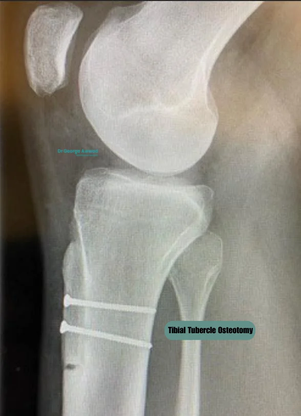
Tibial Tubercle Osteotomy
Realigning the kneecap to reduce pain and prevent dislocations
Tibial Tubercle Osteotomy (TTO) is a surgical procedure used to realign the patella (kneecap) in order to improve knee stability, relieve pain, and prevent recurrent dislocations. The tibial tubercle is the bony prominence on the upper front part of the shinbone (tibia), where the patellar tendon attaches. In some patients, especially those with patellofemoral instability, maltracking, or alignment issues, the position of this bone contributes to knee pain and dysfunction.
TTO involves cutting and repositioning the tibial tubercle to change the line of pull of the patellar tendon, helping to centralise the kneecap within the groove of the femur during movement. It is often performed alongside other procedures such as cartilage restoration or patellofemoral joint stabilisation, depending on the patient’s individual anatomy and underlying issues.
This page provides an in-depth overview of TTO surgery, including when it may be recommended, what to expect before and after surgery, and how Dr George Awwad uses this approach as part of a personalised treatment plan to preserve knee function and prevent future complications.
What is a tibial tubercle osteotomy (TTO)?
A tibial tubercle osteotomy (TTO) is a surgical procedure that aims to correct the alignment of the patella (kneecap) by repositioning a section of bone at the top of the shinbone (tibia) called the tibial tubercle. This small bony prominence is where the patellar tendon attaches, playing a key role in the movement and stability of the kneecap.
When the patella does not track properly in the groove of the femur (thighbone), it can lead to pain, instability, and damage to the cartilage underneath the kneecap. TTO helps to address this by shifting the tibial tubercle in one or more directions, most commonly medially (inwards), anteriorly (forwards), or distally (downwards), to improve patellar alignment and reduce stress on the joint. This realignment procedure can be performed on its own or in combination with other surgeries such as medial patellofemoral ligament (MPFL) reconstruction or cartilage repair, depending on the individual’s specific condition. It is commonly used as part of the treatment plan for patellofemoral instability, maltracking, or chondral damage related to kneecap movement.
The ultimate goal of a TTO is to help stabilise the kneecap, relieve pain, improve knee function, and prevent further joint deterioration, particularly in active individuals or younger patients seeking to delay or avoid more extensive joint replacement procedures.
When might a TTO be recommended?
A tibial tubercle osteotomy (TTO) may be recommended when symptoms such as patellar instability, maltracking, or anterior knee pain are linked to abnormal positioning or alignment of the tibial tubercle, the bony prominence on the shinbone (tibia) where the patellar tendon attaches. This procedure aims to correct the alignment of the patella (kneecap) by repositioning the tibial tubercle, helping improve joint function and reduce mechanical stress within the knee.
TTO is often considered as part of a joint-preserving strategy, particularly for younger or active patients who have not responded to non-surgical treatment. It may also be recommended in combination with other surgical procedures, such as medial patellofemoral ligament (MPFL) reconstruction, to address patellofemoral instability more comprehensively.
Common scenarios where TTO may be considered:
Recurrent patellar dislocations or subluxations
Patellar maltracking
Patellofemoral joint overload or cartilage wear
Abnormal biomechanics or alignment
Failed previous soft tissue procedures
Who might be suitable for TTO surgery?
Tibial tubercle osteotomy (TTO) may be suitable for patients who experience ongoing patellofemoral joint issues such as instability, maltracking, or anterior knee pain and have underlying alignment abnormalities that contribute to their symptoms. TTO is often considered in younger or active patients, particularly when conservative treatments such as physiotherapy, activity modification, or bracing have not provided adequate symptom relief. The goal of the procedure is to correct the position of the tibial tubercle to improve patellar tracking, reduce mechanical overload, and support long-term joint preservation.
You may be a candidate for TTO if:
You have recurrent patellar dislocation or subluxation
You experience persistent patellofemoral pain or maltracking
You have focal cartilage wear or early patellofemoral arthritis
You have anatomical factors contributing to instability
You are physically active and motivated to pursue rehabilitation
You have failed prior soft tissue procedures
During your consultation, Dr George Awwad will perform a detailed clinical assessment and review imaging, including X-rays, CT scans, or MRI, to evaluate your alignment and joint condition. Measurements such as the TT–TG distance, patellar height (Caton–Deschamps Index), and trochlear shape will help determine whether TTO is appropriate and whether it should be combined with other procedures such as MPFL reconstruction or cartilage restoration. Dr Awwad will also consider your goals, age, activity level, and overall knee health to provide individualised surgical recommendations.
Who may not be suitable for this procedure?
TTO may not be suitable if you have:
Advanced patellofemoral arthritis
Diffuse or tricompartmental knee arthritis
Poor bone quality or osteoporosis
Open growth plates (skeletal immaturity)
Infection or active inflammatory joint disease
Unwillingness or inability to follow rehabilitation protocols
Tibial tubercle realignment techniques: medialisation, anteriorisation, distalisation
1. Medialisation
- Purpose: To correct lateral (outer) patellar tracking or recurrent dislocation.
- How it helps: Medialisation repositions the pull of the patellar tendon, helping to keep the kneecap more centred within the femoral groove during movement.
- Often used for: Patients with patellar instability, lateral maltracking, or abnormal tibial tubercle–trochlear groove (TT–TG) distance.
2. Anteriorisation
This technique shifts the tubercle forward (anteriorly).
- Purpose: To reduce pressure on the undersurface of the kneecap.
- How it helps: Anteriorisation unloads the patellofemoral joint, which may reduce pain and inflammation in patients with early cartilage wear or chondral defects.
- Often used for: Individuals with patellofemoral pain syndrome, chondromalacia patellae, or focal cartilage defects.
3. Distalisation
Distalisation lowers the tibial tubercle, moving it downward toward the foot.
- Purpose: To correct a high-riding patella (patella alta), which can impair engagement with the trochlear groove.
- How it helps: This improves the patella’s contact with the femur during knee flexion, supporting better joint stability and function.
- Often used for: Patients with patella alta and recurrent dislocations.
Tailoring the approach to your anatomy
In many cases, a combination of movements such as anteromedialisation (forward and inward) may be used to achieve optimal alignment and joint load distribution. Dr George Awwad uses detailed imaging and measurements, including TT–TG distance and patella height, to plan the direction and degree of adjustment that best matches your knee’s anatomy and surgical goals. This precise, personalised approach supports better outcomes in stability, pain relief, and long-term joint preservation.
TTO vs MPFL Reconstruction vs Lateral Release: how do they differ?
1. Tibial Tubercle Osteotomy (TTO)
- What it does: Realigns the patellar tendon by surgically repositioning the bony attachment on the tibia (shin bone).
- Target area: Bony structures of the lower knee, specifically the tibial tubercle.
- Best suited for: Patients with:
- Abnormal TT–TG distance
- Patella alta (high-riding patella)
- Lateral maltracking
- Patellofemoral overload or cartilage wear
- Goal: Improve patella alignment and reduce joint loading by adjusting how the kneecap tracks through the femoral groove.
2. MPFL Reconstruction
- What it does: Reconstructs the medial patellofemoral ligament, which helps prevent the patella from dislocating laterally (outwards).
- Target area: Soft tissue on the inner (medial) side of the knee.
- Best suited for: Patients with:
- Recurrent patellar dislocations
- MPFL tears or laxity
- Normal patella height and bony alignment
- Goal: Restore soft tissue restraint to stabilise the kneecap, especially in early flexion.
3. Lateral Release
- What it does: Involves releasing tight soft tissues on the outer (lateral) side of the patella.
- Target area: Lateral retinaculum, a fibrous band that can pull the patella outward.
- Best suited for: Selected cases with:
- Lateral tilt of the patella
- Isolated soft tissue tightness
- Goal: Relieve lateral tension to improve patella tracking and reduce pain.
Note: Lateral release is less commonly performed in isolation today, as it may be insufficient on its own and can lead to instability if overused. It is sometimes performed in combination with TTO or MPFL reconstruction.
Selecting the right procedure
- TTO + MPFL reconstruction (for instability with bony malalignment)
- TTO + lateral release (for severe lateral patella tilt and tightness)
What happens during the tibial tubercle osteotomy surgery?
Below is an overview of the key steps involved in the procedure:
1. Anaesthesia and positioning
2. Surgical incision
3. Cutting and mobilisation of the tibial tubercle
4. Repositioning and fixation
5. Additional procedures (if needed)
- MPFL reconstruction (to restore soft tissue stability)
- Lateral release or lengthening (to reduce outer pull on the kneecap)
- Cartilage repair or debridement (if damage is found inside the joint)
6. Wound closure and dressing
7. Post-operative care and monitoring
What is the recovery process after TTO?
Recovery after a Tibial Tubercle Osteotomy (TTO) is a gradual, structured process designed to protect the surgical site, optimise healing, restore strength and function, and eventually support a safe return to daily and sporting activities. Recovery timelines can vary depending on your anatomy, the extent of the procedure, and whether additional procedures (such as MPFL reconstruction or lateral release) were performed. Dr George Awwad follows a detailed rehabilitation protocol that includes specific phases and guidance for weight-bearing, bracing, physiotherapy, and return to activity.
Initial recovery: first 1–3 weeks
- Weight-bearing: You will typically begin with touch weight-bearing using crutches.
- Knee brace: A hinged knee brace is worn at all times (except during hygiene routines) and is initially set to allow 0–30 degrees of movement.
- Pain management: You’ll be prescribed pain relief and advised to use ice packs and leg elevation regularly to help manage swelling and discomfort.
- Compression: White compression stockings may be used for the first 2 weeks to reduce the risk of blood clots.
- Dressings: Bulky surgical dressings are removed the day after surgery, with smaller adhesive dressings kept intact. A wound check is arranged within 1–2 weeks.
- Physiotherapy: Early, supervised physiotherapy begins immediately to minimise stiffness and promote safe movement. Exercises may include:
- Heel slides
- Isometric quadriceps contractions
- Gentle assisted flexion
- Calf and hamstring stretches
Weeks 4–6: Improving mobility and range of motion
- Weight-bearing: Progression to partial weight-bearing with crutches.
- Brace settings: The knee brace is adjusted to allow 0–45 degrees of movement.
- Physiotherapy: Continues with a focus on increasing range of motion, maintaining flexibility, and improving muscle control. New exercises may include:
- Prone assisted knee flexion
- Strengthening of calf and hamstring muscles
- Stretching and neuromuscular stimulation (as needed)
Weeks 6–12: Building strength and movement
- Imaging review: Before progressing, X-rays are used to confirm healing of the osteotomy site.
- Weight-bearing: You may begin weight-bearing as tolerated (WBAT) and gradually wean off crutches as strength allows.
- Brace: May be worn with increasing flexibility, eventually allowing 0–90 degrees of motion, then weaned off based on strength and stability.
- Exercise progression: You may begin using:
- Stationary bike
- Elliptical trainer
- Stairmaster (short steps)
- Pool walking (from 8 weeks)
Months 3–6: Return to function and low-impact activity
- Criteria for progression: Minimal swelling, no pain, and knee flexion of at least 120 degrees.
- Goals: Improve lower limb strength, restore balance and coordination, and prepare for return to work or sport.
- Exercises may include:
- Knee extensions
- Eccentric strengthening
- Cardiovascular conditioning (bike, elliptical, swimming)
- Proprioceptive training
Months 6–9: Return to higher-level activity
- Criteria for progression: No patellofemoral pain or swelling, near-normal range of motion, and 70% strength compared to the unaffected leg.
- Activities may now include:
- Light jogging
- Plyometric exercises
- Sport-specific drills (e.g. agility ladders, shuttle runs, cutting movements)
9+ Months: Return to sport or demanding physical activity
- Full return to sport or high-demand work is typically permitted around 12 months after surgery, pending medical clearance from Dr Awwad.
- You will continue a maintenance program tailored to your goals and physical condition.
Ongoing physiotherapy support
Physiotherapy plays a key role throughout each recovery phase. Dr Awwad recommends 6–12 months of supervised rehabilitation, which will be adjusted based on your progress and goals
What are the potential risks or complications of TTO?
As with any orthopaedic procedure, tibial tubercle osteotomy (TTO) carries certain risks and potential complications. While many patients recover well with appropriate rehabilitation, it’s important to be aware of possible challenges that may arise before, during, or after surgery. Dr George Awwad will discuss these risks with you during your consultation and answer any questions you may have as part of the informed consent process.
General surgical risks
Infection
Bleeding or haematoma
Blood clots (deep vein thrombosis or pulmonary embolism)
Wound healing problems
Risks specific to tibial tubercle osteotomy
Non-union or delayed union
Fracture
Loss of fixation
Hardware irritation
Overcorrection or undercorrection
Patella baja or alta
Functional complications
- Persistent pain or stiffness: Some patients may experience ongoing discomfort or reduced knee motion after surgery.
- Reduced strength or weakness: Temporary quadriceps weakness is common but usually improves with physiotherapy.
- Nerve or vessel injury: Although rare, there is a small risk of injury to surrounding nerves or blood vessels.
Need for further surgery
In some cases, further surgery may be needed. This may include:
- Hardware removal due to discomfort
- Arthroscopy or revision realignment procedures if symptoms persist
- Progression to patellofemoral or total knee replacement if underlying joint degeneration advances
Tibial Tubercle Osteotomy FAQs
What is a tibial tubercle osteotomy and why is it done?
Tibial tubercle osteotomy (TTO) is a surgical procedure that repositions the bony attachment of the patellar tendon to correct misalignment of the kneecap (patella). It is often recommended for patients with recurrent patellar instability, maltracking, or patellofemoral joint pain due to abnormal biomechanics.
How is the bone fixed in place during a TTO?
During the procedure, the tibial tubercle is carefully cut and moved to a new position (typically medialised, anteriorised, or distalised). It is then securely fixed in place using surgical screws. These screws are usually left in permanently but can be removed if they cause irritation once the bone has healed.
Does the bone grow back together after TTO surgery?
Yes, the bone typically heals over a period of 6 to 12 weeks. During this time, the bone gradually forms a solid union in the new position. Weightbearing is often limited initially to protect the healing site, and serial imaging may be used to confirm bone healing.
Will I need to wear a brace after TTO surgery?
Yes. Most patients are fitted with a knee brace immediately after surgery. This brace helps stabilise the knee, limits unwanted motion, and protects the osteotomy site while the bone heals. Your physiotherapist will guide you on how and when to adjust or remove the brace as you progress through rehabilitation.
How long will I be on crutches after a tibial tubercle osteotomy?
Can a TTO delay or prevent the need for knee replacement?
In certain cases, yes. When combined with other procedures (such as cartilage repair or realignment), a TTO can reduce abnormal loading on the patellofemoral joint and may help preserve knee function, potentially delaying the need for partial or total knee replacement in younger patients with isolated patellofemoral arthritis or malalignment.
When can I return to work after TTO surgery?
- Sedentary/desk-based work: You may be able to return within 2–4 weeks.
- Light physical work: May require 6–8 weeks off.
- Heavy manual work: A longer recovery period of 3–4 months may be required.
Dr Awwad will provide personalised guidance based on your role and recovery progress.
When can I drive after a TTO?
Driving is typically not recommended until you can safely bear weight, have sufficient range of motion, and can control the pedals without pain or delay. This usually occurs around 6–8 weeks after surgery, depending on whether your operative leg is the left or right side and whether your vehicle has manual transmission.
When can I return to sport after TTO surgery?
- Low-impact exercise (e.g. cycling, swimming): Usually allowed after 3 months.
- Running or agility-based sports: May require 5–6 months or longer. A gradual, supervised return is essential and guided by your physiotherapist.
What role does physiotherapy play after tibial tubercle osteotomy?
- Early phase: swelling control, range of motion, and quadriceps activation.
- Intermediate phase: increased loading, gait retraining, and functional exercises.
- Late phase: sport-specific training, agility, and strength restoration.